Continuous Suture Device for Gastrointestinal Endoscope
In recent year, NOTES(Natural Orifice Transluminal Endoscopic Surgery) have been an active research area of Engineering and Medical science. It doesn't make incisions on the body and leave scars because it performs abdominal operations with a multi-channel endoscope passed through a natural orifice (mouth, urethra, anus, and vagina). Bariatric surgery for treating obesity and GERD (gastro-esophageal reflux disease) surgery are the good examples applicable NOTES. One of the important issues of these endoscopic surgeries is suturing. Unfortunately, conventional endoscopic instruments can perform only few stitches. We are developing Gastrointestinal Endoscope which is capable of suturing continuously.
The Structure of continuous suture device
The continuous suture device is designed to be installed in a conventional gastrointestinal endoscope, and is composed of three parts, as shown in Fig. 2: needle, tube, and handle. The needle component consists of the 18-gauge needle itself, beads, and a thread. It penetrates and holds the tissues like sewing. The tube component consists of a protective tube covering the exterior, the needle transfer tube, the bead transfer tube, and the thread. The handle component enables relative movement between the other two components. Its control should be easy.

Fig 1. Structure of continuous suture device
A side suction cap is designed to exert suction on tissue. The tissue is sucked into the cap after closing the elastic hinge door that prevents failure of the semi-vacuum due to air inflow from the outside during the suction. The needle is protected by the side suction cap before insertion into the tissue during the operation.
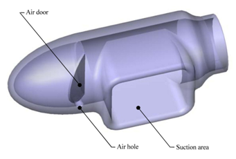
Fig 2. Side suction cap
Surgical Procedure with the continuous suture device
(a) Part of the stomach tissue is sucked into the side suction cap.
(b) The needle penetrates the sucked-in tissue, and then four or five stitch beads are advanced.
(c) The advanced beads form a circular loop when the thread is pulled.
(d) One stitch is completed

Fig 3. Schematic view of the surgical procedure
Experiments
To validate the device experiments are carried some times. Animal experience using pig are planned.
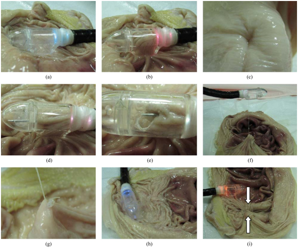
Fig 4. View of experiment of continuous suture device for validation using extracted stomachs
References
- Bae, K., Jung, K., Chu, B., Hong, D. H., Chun, H. J., Kim, Y. and Keum, B., "Development of a novel successive suturing instrument for an endoscope using beads and a side suction cap", Int. J. Precis. Eng. Manuf., 10(2), 97-103, 2009.